
Basic Ventriculostomy Care: Practical Points for Non-Neurosurgeons
By Intouch Sopchokchai, M.D.
What Is a Ventriculostomy?
A ventriculostomy is a procedure that places a drainage catheter into the brain’s ventricle to drain cerebrospinal fluid (CSF). It is indicated to monitor intracranial pressure (ICP) and to help lower elevated ICP by draining CSF through the catheter. The height at which the ventriculostomy is set controls the flow of CSF in and out — and therefore intracranial pressure — based on the neurosurgeon’s assessment and order.
Setup
“Raising” the ventriculostomy (measured in cm above a reference level) means that if the patient’s intracranial pressure exceeds the set height, CSF will flow out to rebalance the pressure. Setting the ventriculostomy lower therefore means CSF drains out more easily than setting it higher.
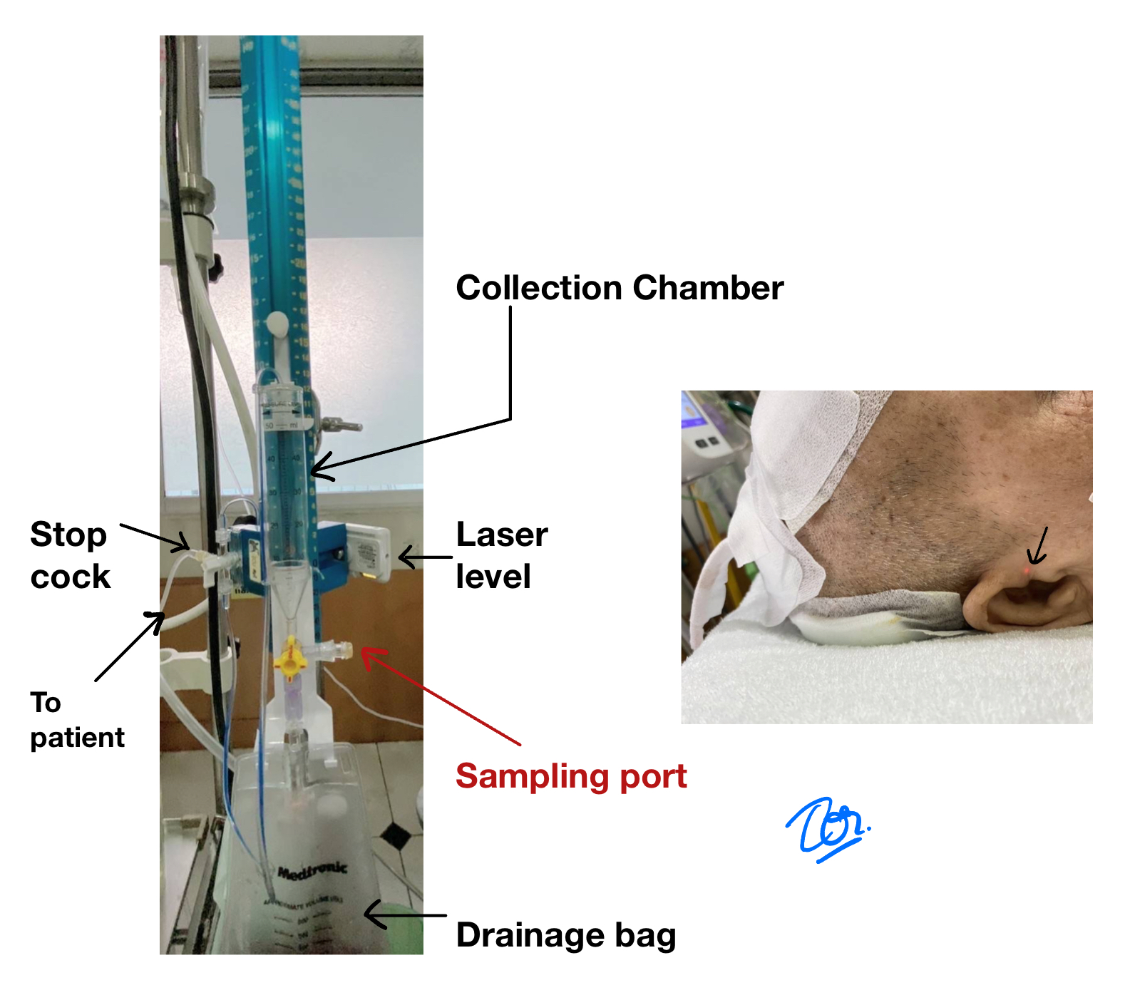
The height should be measured from the patient’s reference point (typically the external auditory meatus) to the highest point of the system. For example, an order to “raise the ventriculostomy 20 cm” means the highest point of the tubing should sit 20 cm above the patient’s ear canal — how far the system sits horizontally from the patient doesn’t matter.
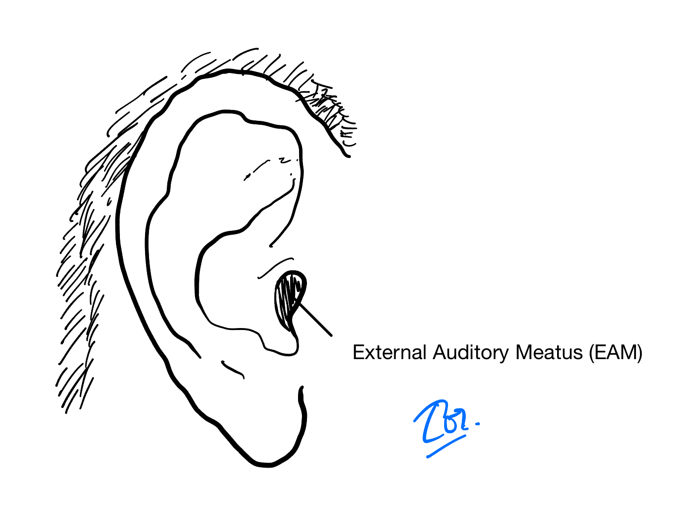
The precise reference point is the external auditory meatus when the patient is supine, and the midpoint of the head (the point between the eyebrows) when the patient is lying on their side.
Because the reference point is tied to the patient’s ear, the ventriculostomy height must be re-measured and re-set every time the patient’s head height changes — for example, when the bed is raised or lowered, or when the patient slides down an inclined bed.
Precautions
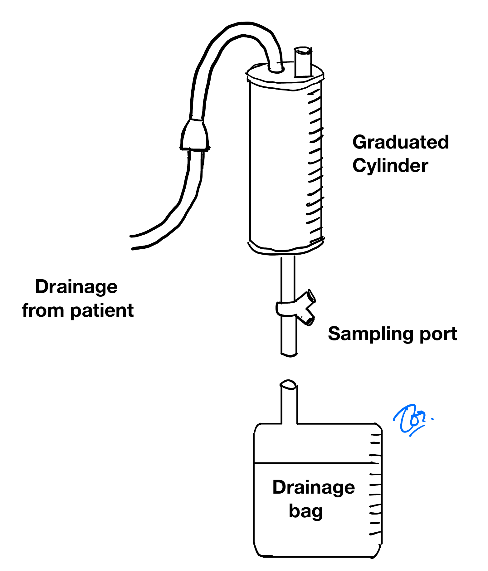
Height is measured from the highest point of the system, which differs between a commercial set and an extension tube:
- Commercial set: measured from the stopcock.
- Extension tube: measured at the highest point before the tubing curves down into the collection cylinder.
Assessing Ventriculostomy Function
A well-functioning ventriculostomy should:
- Fluctuate with the cardiac and respiratory cycle.
- Flow back toward the head when the system is raised, and away from the head when lowered.
If the ventriculostomy does not fluctuate or shows no inflow/outflow, the catheter may be obstructed or displaced — notify the neurosurgeon to consider irrigation or additional imaging.
An increase or decrease in CSF output does not by itself indicate whether the catheter is blocked. It’s normal for CSF to stop draining once intracranial pressure equals the external pressure, just as it’s normal for CSF to drain heavily while intracranial pressure remains elevated.
If the fluid column still fluctuates but CSF cannot flow in or out, the catheter tip may be lodged in brain parenchyma, fluctuating with parenchymal pulsation — this should be assessed clinically and/or with imaging. If CSF flows but does not fluctuate, the catheter tip may be poorly positioned; whether to revise it depends on the clinical situation.
If a patient’s level of consciousness remains altered after ventriculostomy placement, consider the following:
CSF Underdrainage
Signs of persistent increased intracranial pressure:
- Lethargy
- Headache
- Vomiting
- Bulging fontanelle (in infants)
- CT: ventricles remain enlarged
Possible causes:
- Catheter tip malpositioned
- Ventriculostomy system obstructed
- Ventriculostomy set too high, draining insufficient CSF
CSF Overdrainage
Signs suggestive of intracranial hypotension:
- Headache
- Tachycardia
- Vomiting
- Blurred vision
- Sunken fontanelle, overlapping sutures (in infants)
- CT: slit ventricles
Possible causes:
- CSF released too quickly and in too great a volume — consider lying the patient flat, clamping the ventriculostomy, and giving IV hydration.
Other contributing factors:
- Intracranial hemorrhage
- Seizure
- Infection
- Other metabolic causes
Collecting CSF and Administering Medication via Ventriculostomy
Most ventriculostomy systems include a three-way stopcock (T-way stopcock) in the line, allowing CSF to be collected for testing. CSF should not be withdrawn with a syringe — the catheter tip may sit close to brain tissue or the choroid plexus, and negative-pressure suction risks aspirating brain tissue, causing injury and bleeding. Instead, open the T-way stopcock and allow fluid to drain on its own, using sterile technique every time. If unsure, notify the neurosurgeon.
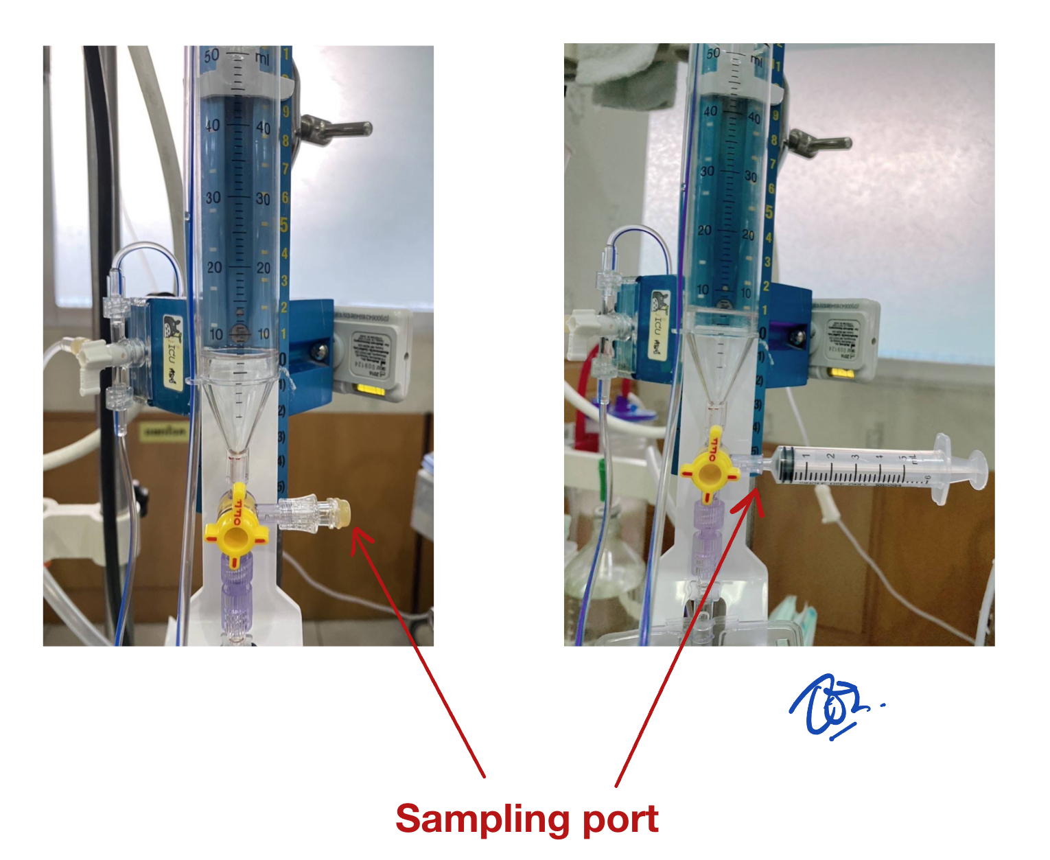
Medication can also be given through the ventriculostomy, at the neurosurgeon’s discretion. If needed, medication can be given through the T-way stopcock, pushed in as slowly as possible with a syringe, followed by a slow normal saline flush to clear the line. Before administering medication, drain an equivalent volume of CSF to accommodate the medication and saline being added, using the technique described above.
Other General Care
Clamp the ventriculostomy during patient activity, since movement or straining raises intracranial pressure and can cause excess CSF drainage. Activities to watch for include:
- Bathing / wiping down the patient
- Transferring or moving the patient
- Suctioning secretions
If the wound site is weeping, consider opening the dressing to assess for CSF leakage or wound complications. Avoid opening the dressing more often than necessary — some recommendations suggest re-dressing every 2–3 days, while others suggest a single dressing change on postoperative day 5–7.
Since meningitis is commonly seen after postoperative day 5–7, reassess whether the ventriculostomy is still indicated. If not, consider removal; if CSF drainage is still needed, consider replacing it at a new site. When removing a ventriculostomy, always suture the drain site closed. Antibiotic prophylaxis is not required.
In certain cases — such as a ruptured aneurysm — if the ventriculostomy output suddenly darkens with fresh-looking blood and drains rapidly, accompanied by a sudden drop in Glasgow Coma Score, this may indicate aneurysm re-rupture. Notify the patient’s neurosurgeon immediately.
This article was originally published on the department's previous website.